Figure 3 from Stimulated Emission Depletion (STED) Microscopy: from Theory to Practice
Por um escritor misterioso
Last updated 24 fevereiro 2025

Figure 3. Fluorescence depletion of two common dyes in STED microscopy, Atto647N (black, diamonds) and Atto655 (red, circles), as a function of the depletion laser intensity. Error bars for Atto647N appear smaller than the point size of the average value. - "Stimulated Emission Depletion (STED) Microscopy: from Theory to Practice"

Stochastic optical reconstruction microscopy (STORM) in comparison

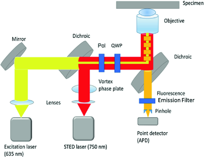
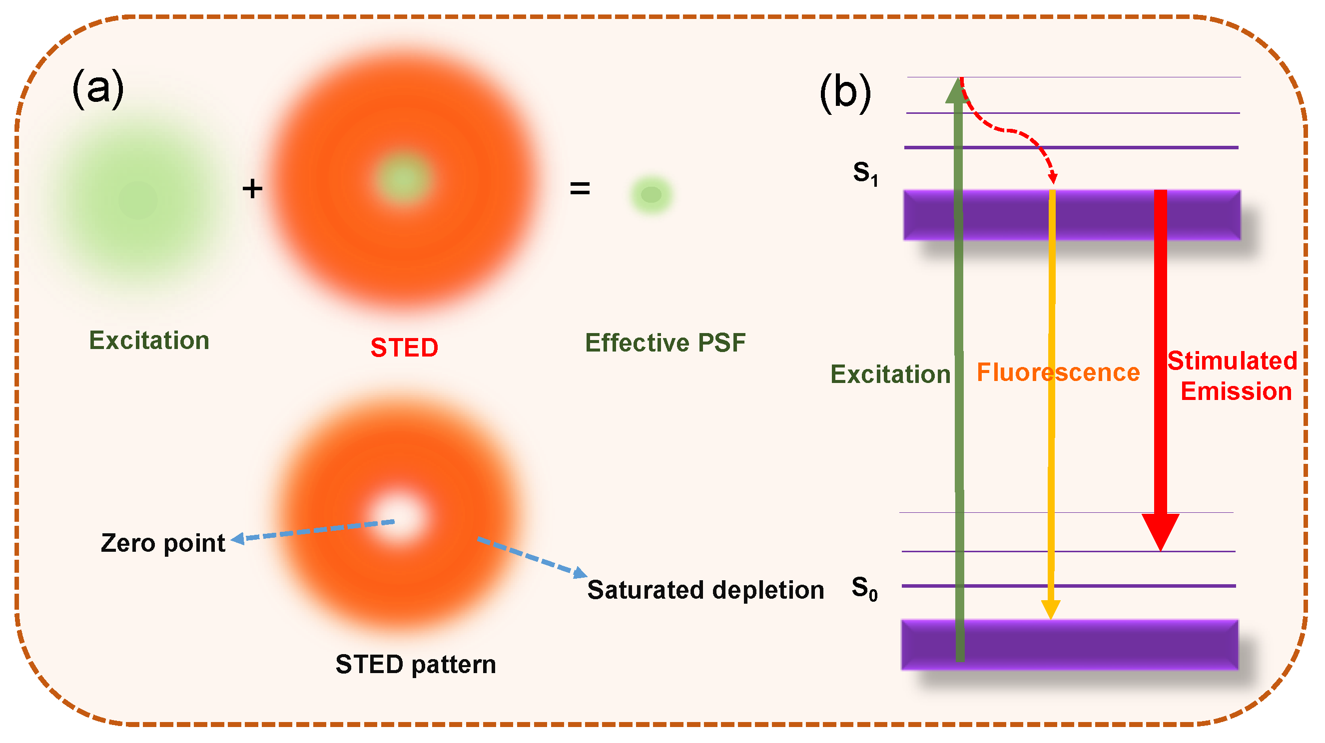
Stimulated Emission Depletion Microscopy

Building a fast scanning stimulated emission depletion microscope

PDF) Stimulated Emission Depletion (STED) Microscopy: from Theory

Principle of STED microscopy. The red depletion beam is phase
Two-photon excitation and stimulated emission depletion by a

Ultralow power demand in fluorescence nanoscopy with digitally

Stimulated Emission Depletion Microscopy

Stimulated emission depletion (STED) nanoscopy of a fluorescent

Nanoparticle-Assisted Stimulated-Emission-Depletion Nanoscopy
Stimulated Emission Depletion Microscopy and Related Techniques

3 Principles underlying stimulated emission depletion (STED
Materials, Free Full-Text
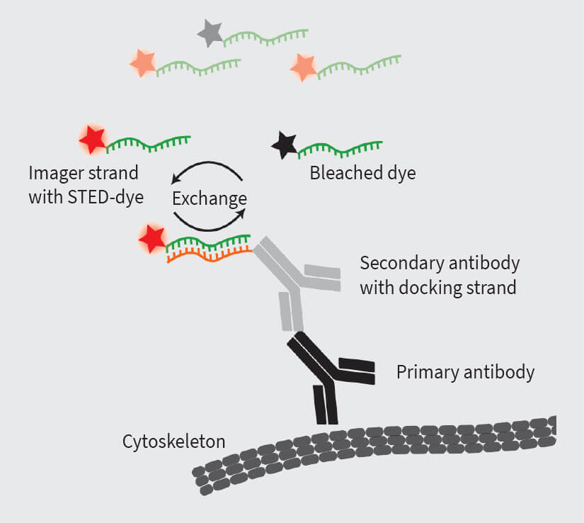
STED-PAINT for high-performance superresolution
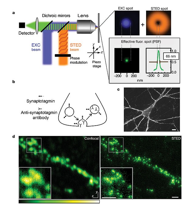
STED microscopy reveals that synaptotagmin remains clustered after
Recomendado para você
-
Peartree Practice24 fevereiro 2025
-
 Chessel Avenue, Southampton, SO19 4DY 4 bed detached house - £650,00024 fevereiro 2025
 -
Victor Rodao Gonzalez - First contact Physiotherapist - PEARTREE practice24 fevereiro 2025
-
 Peartree Practice24 fevereiro 2025
 -
 The Hub Appointments24 fevereiro 2025
 -
 Montessori Toddler Discipline : Modern Stress-Free Parenting Guide with Practical Approach and Strategies to Tame Tantrums, Conflicts and Raise a24 fevereiro 2025
 -
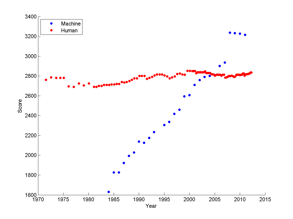 CS440 Lectures24 fevereiro 2025
 -
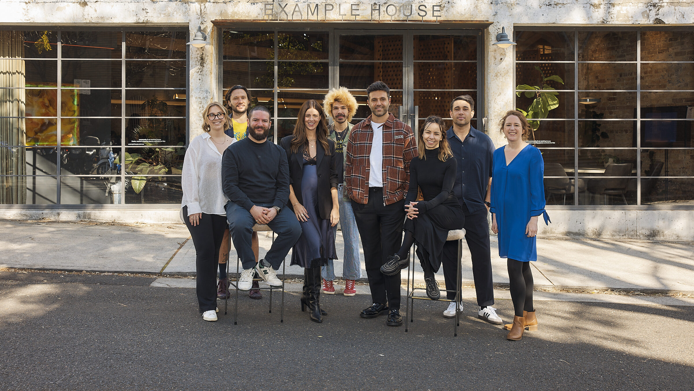 Example launches new positioning, makes several new hires24 fevereiro 2025
 -
 General Practice Transformation Champions: Improving Access to General Practice24 fevereiro 2025
 -
 Plants, Free Full-Text24 fevereiro 2025


você pode gostar
-
10 Worst Moments in Doors Roblox Part 3, Robstix Wiki24 fevereiro 2025
-
 Rex Orange County Teases New Song on TikTok24 fevereiro 2025
 -
 COMO DESENHAR PAPER DUCK +PIJAMA+ ROUPINHAS #paperducks #paperdolls #bonecadepapel #shorts24 fevereiro 2025
 -
 Vasco x Fluminense: onde assistir, horário e escalações do jogo pelo Brasileirão24 fevereiro 2025
 -
 Cozinhar bolo de morango na App Store24 fevereiro 2025
 -
 Panda bonito quebra-cabeça - Jogo aprendizagem 3D para quebra-cabeças infantis,Molduras para fotos Jogos Panda Decoração casa para pare, janela para24 fevereiro 2025
 -
 18 WHEELER 3D jogo online gratuito em24 fevereiro 2025
 -
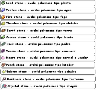 Pedras da evolução (stones): O que são? Como conseguir? - Dentro24 fevereiro 2025
 -
 Prisma Instrumentos Odontológicos24 fevereiro 2025
 -
 🔥 999+ Stylish Boy DP For Instagram & Whatsapp Profile Pic 202324 fevereiro 2025