Electron microscopy and calorimetry of proteins in supercooled
Por um escritor misterioso
Last updated 24 fevereiro 2025
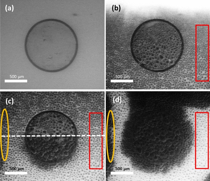
Active site identification on feldspar from freeze-thaw
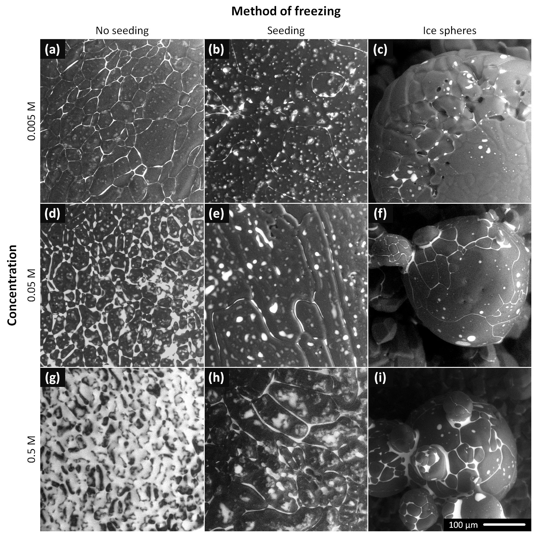
TC - The morphology of ice and liquid brine in an environmental
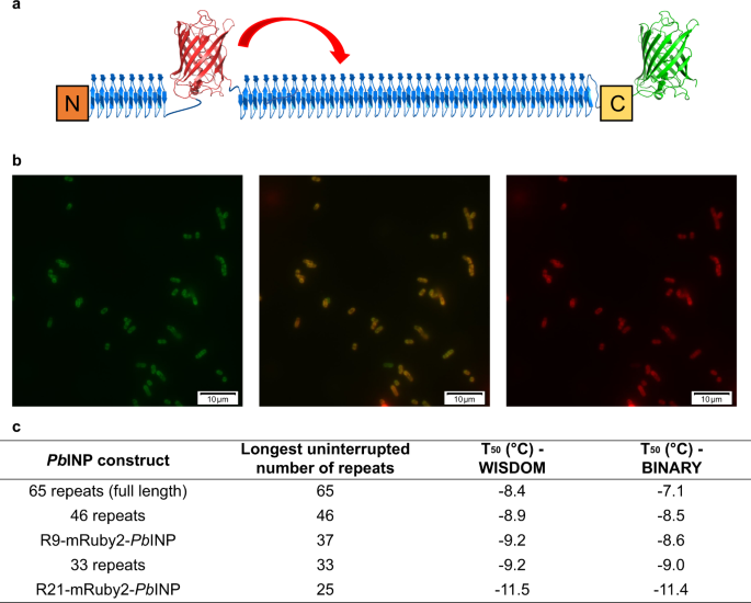
Water-organizing motif continuity is critical for potent ice

Cumulative nucleus spectra for multiple size fractions of soil

a) Amide I band (1630 cm À1 ) of the infrared spectra of e-PLL, pH

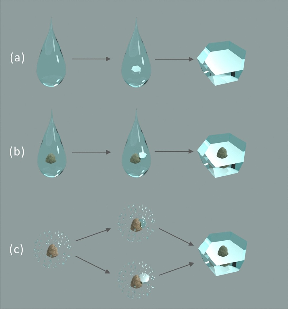
a) Left: p-T diagram showing the extent of supersaturation
Electron microscopy and calorimetry of proteins in supercooled

Temperature Derivative Fluorescence Spectroscopy as a Tool to

Structure of spruce budworm antifreeze protein.a, Stereoview of C
Recomendado para você
-
 A Verified Bot that generates Visual Random State Scrambles! (more24 fevereiro 2025
 -
John Walters Certified Arborist24 fevereiro 2025
-
 WCA 2021 Round Up - WFSA24 fevereiro 2025
 -
 Sustainable land management for improved livelihoods and24 fevereiro 2025
 -
 Ionically and Enzymatically Dual Cross-Linked Oxidized Alginate24 fevereiro 2025
 -
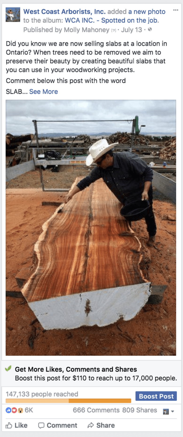 Local Business Sells Out of Wood Using Messenger Bots Case Study24 fevereiro 2025
 -
 A Bot that Generates Regripless Scrambles! (more info in the24 fevereiro 2025
 -
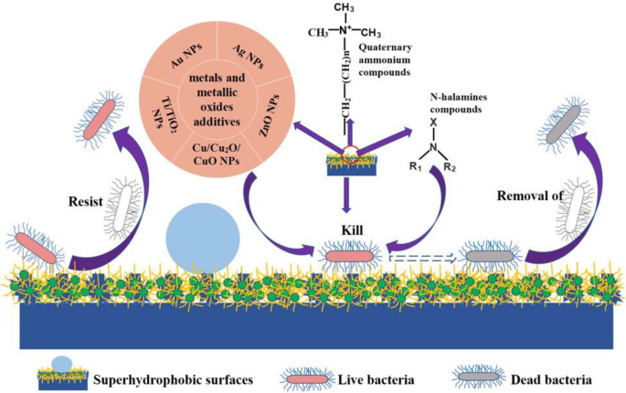 Recent Advances in Superhydrophobic and Antibacterial Cellulose24 fevereiro 2025
 -
 jordan mcann and king arnold|TikTok Search24 fevereiro 2025
 -
 Watercolor Artist Fall 2022 Digital Edition24 fevereiro 2025

você pode gostar
-
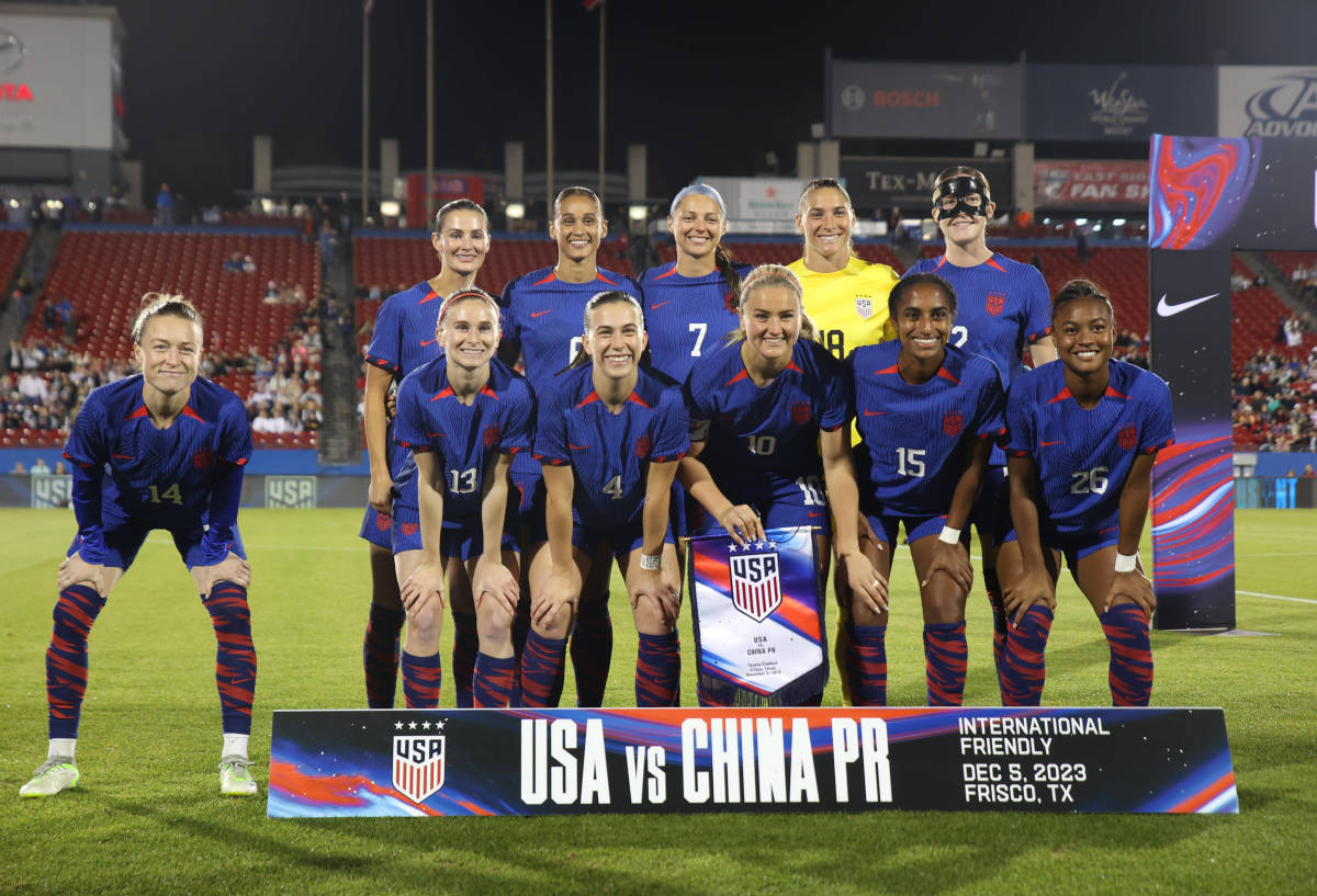 USWNT up to 2nd in FIFA Women's Women's Rankings, Spain 1st - Futbol on FanNation24 fevereiro 2025
 -
 Ajedrez en línea - Juega en línea en SilverGames 🕹️24 fevereiro 2025
 -
 Wallpaper : Kimetsu no Yaiba, Kamado Tanjiro, Kamado Nezuko, anime24 fevereiro 2025
 -
:strip_icc()/i.s3.glbimg.com/v1/AUTH_bc8228b6673f488aa253bbcb03c80ec5/internal_photos/bs/2021/B/p/41SKM5Ski3q7I5e4uGow/fmf-segundona.jpg) FMF divulga tabela da Segunda Divisão do Mineiro 2021; veja 1ª rodada, futebol24 fevereiro 2025
FMF divulga tabela da Segunda Divisão do Mineiro 2021; veja 1ª rodada, futebol24 fevereiro 2025 -
 Roblox Tapping Simulator Rebirth System Scripting Tutorial24 fevereiro 2025
 -
animesvision.biz Traffic Analytics, Ranking Stats & Tech Stack24 fevereiro 2025
-
 Tata Steel Downstream Products Ltd appoints Karan Lakhani as CHRO24 fevereiro 2025
 -
Replying to @psyco648 Rick roll in the weather? Challenge accepted! #r24 fevereiro 2025
-
 Slobodna Dalmacija - Počinje xSTatic graffiti škola: mladi Splićani, javite se, sudjelovanje i svi materijali su besplatni24 fevereiro 2025
 -
 Merlyn é a grande vilã de Nanatsu no Taizai24 fevereiro 2025
