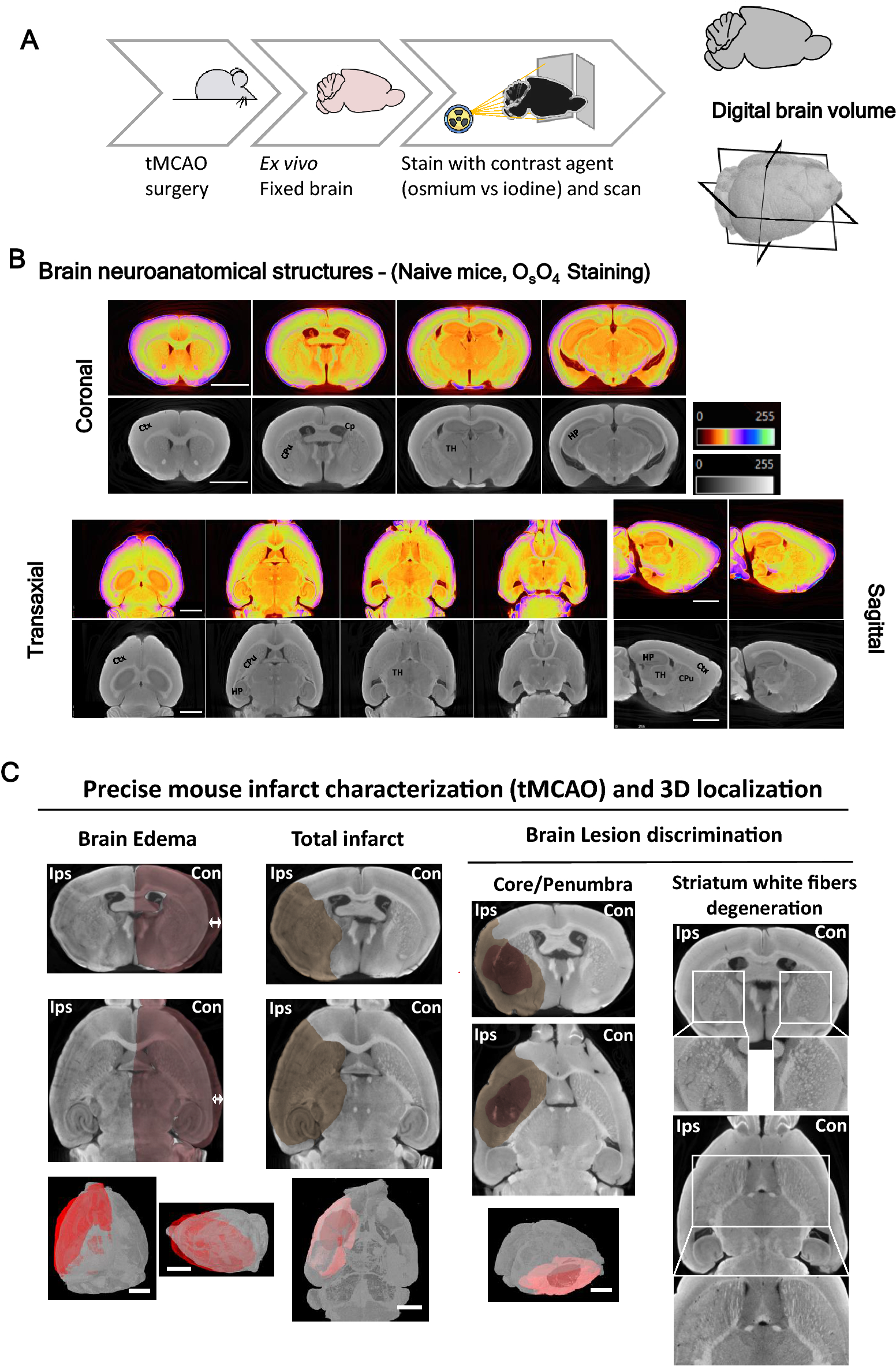
High-resolution micro-CT for 3D infarct characterization and segmentation in mice stroke models
Por um escritor misterioso
Last updated 13 março 2025

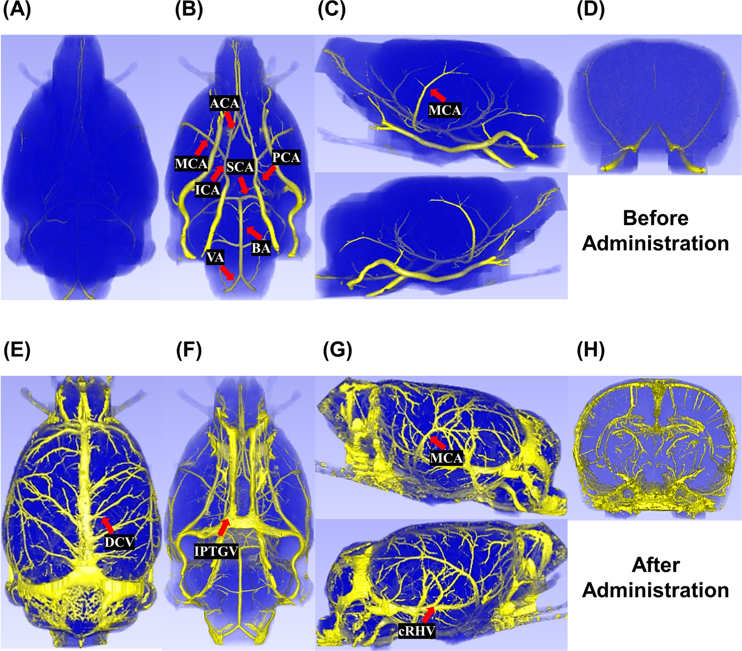
A New Micro–Computed Tomography–Based High-Resolution Blood–Brain Barrier Imaging Technique to Study Ischemic Stroke

WhiceCT compared to in vivo µMRI. (A) Reconstructed images of whiceCT

Micro-CT of rodents: state-of-the-art and future perspectives. - Abstract - Europe PMC
Cells, Free Full-Text

3D micro-CT imaging of the postmortem brain. - Abstract - Europe PMC

PDF) High resolution micro-CT imaging in mice stroke models: from 3D detailed infarct characterization to automatic area segmentation

Identification of anatomical structures in mouse brain in micro CT and
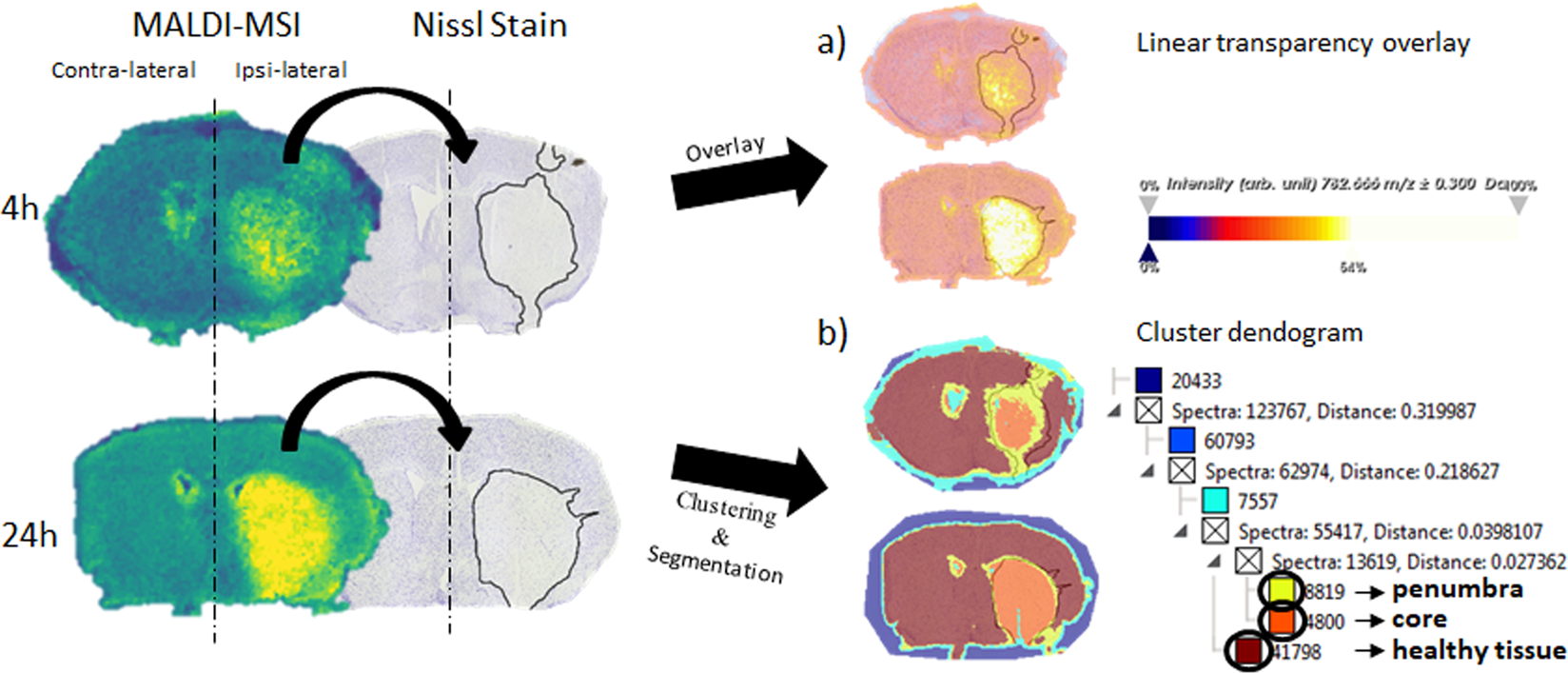
Distinguishing core from penumbra by lipid profiles using Mass Spectrometry Imaging in a transgenic mouse model of ischemic stroke

Complications and Pitfalls in Rat Stroke Models for Middle Cerebral Artery Occlusion

Atlas registration for edema-corrected MRI lesion volume in mouse stroke models - Stefan Koch, Susanne Mueller, Marco Foddis, Thomas Bienert, Dominik von Elverfeldt, Felix Knab, Tracy D Farr, René Bernard, Monika Dopatka

3D visualization and quantification of microvessels in the whole ischemic mouse brain using solvent-based clearing and light sheet microscopy - Erlen Lugo-Hernandez, Anthony Squire, Nina Hagemann, Alexandra Brenzel, Maryam Sardari, Jana Schlechter

Advances in micro-CT imaging of small animals - ScienceDirect

Whole-body µCT upon transcardial perfusion of Lugol's solution after
MRI Visualization of Whole Brain Macro- and Microvascular Remodeling in a Rat Model of Ischemic Stroke: A Pilot Study
Recomendado para você
-
 Best free Websites to test your Mouse Accuracy13 março 2025
 -
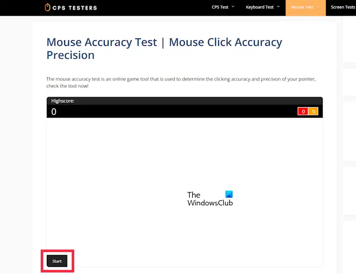
 Mouse Test - How accurate and fast can you move your mouse?13 março 2025
 -
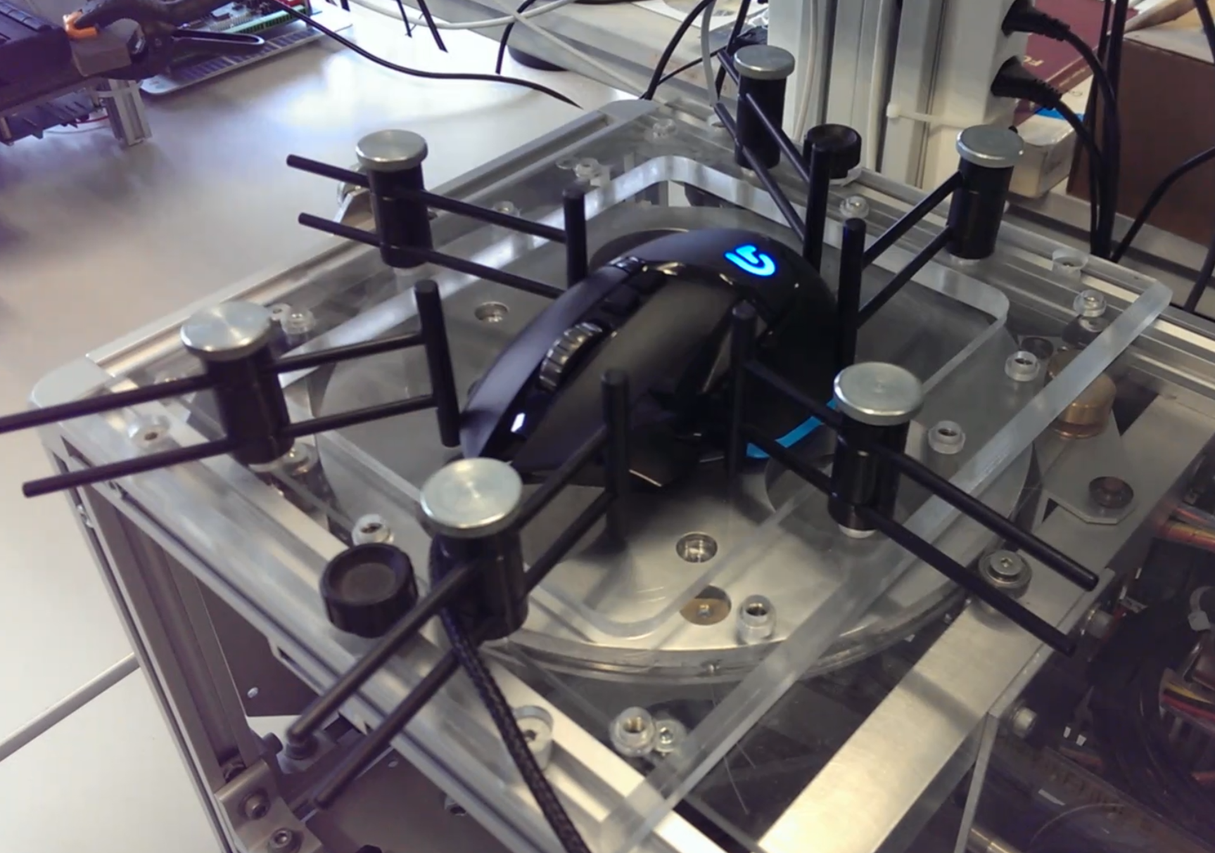
 Mouse Sensor Accuracy Test #1 (G900 vs EC2-A vs PM3360) - Hardware - Mouse Sensitivity Community13 março 2025
 -
 Set up the stereo mapping Stealth 3D mouse—ArcGIS Pro13 março 2025
 -
 ZS-N1, FDM 3D Printed Asymmetric G305 Wireless Mouse Mod, NP-01s inspired : r/MouseReview13 março 2025
 -
 The Testing Facilities - Logitech 2014 Switzerland Tech Day: The Hills Are Alive With The Sound Of Romer G13 março 2025
 -
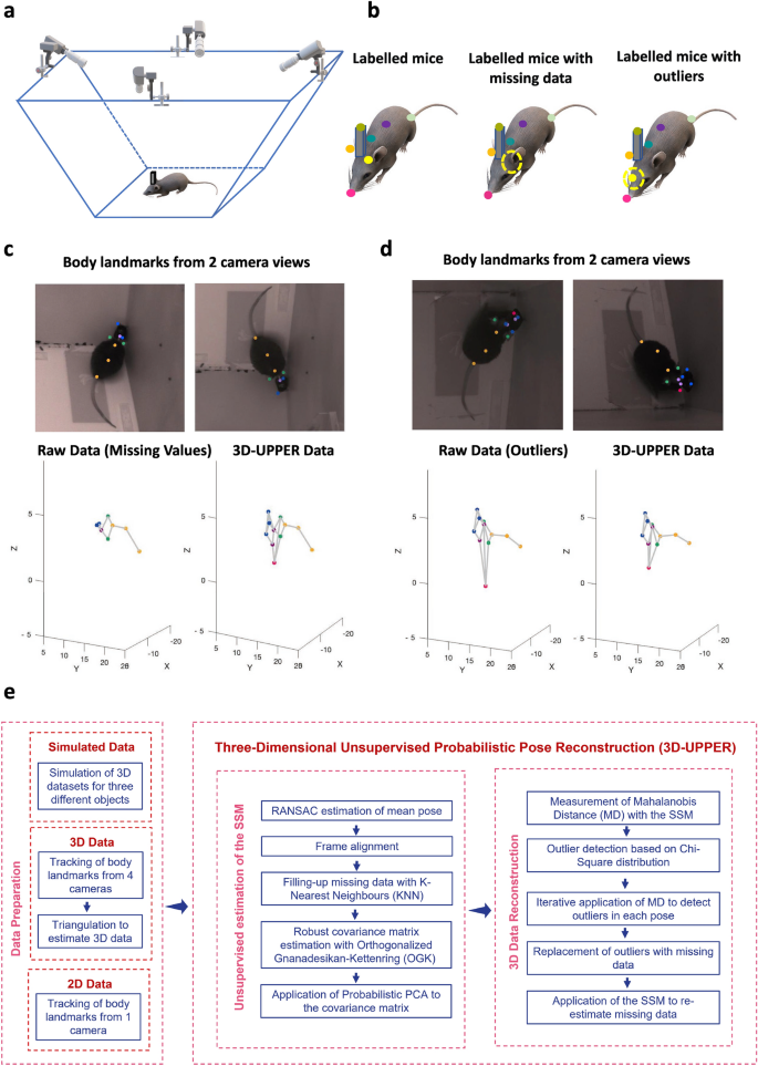 Three-dimensional unsupervised probabilistic pose reconstruction (3D-UPPER) for freely moving animals13 março 2025
 -
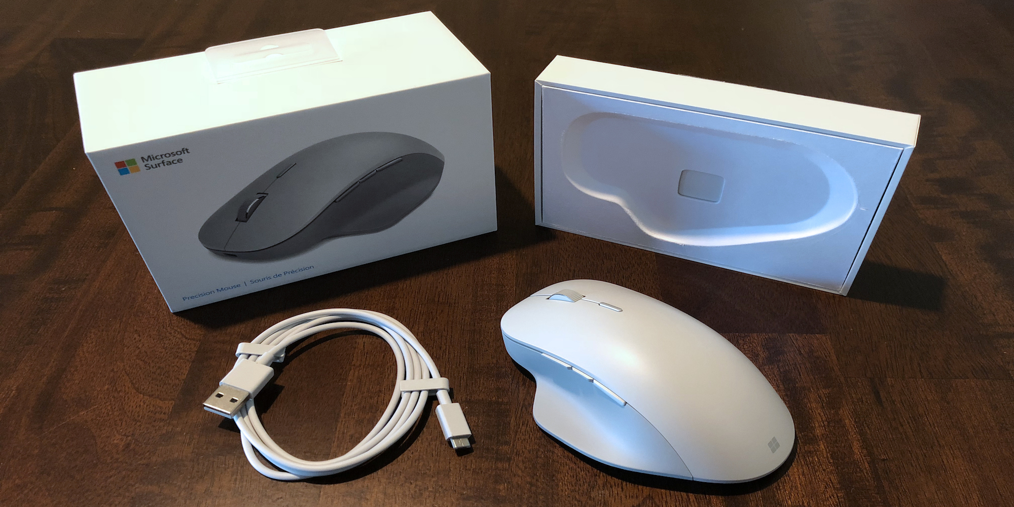 Review: Microsoft Surface Precision Mouse - Thinking Different about Mac functionality - 9to5Mac13 março 2025
 -
Flexible Materials for High-Resolution 3D Printing of Microfluidic Devices with Integrated Droplet Size Regulation13 março 2025
-
 Put 3D Metal Printing Services To The Test, By Making A Watch13 março 2025

você pode gostar
-
 The Grind Outdoors 3 Pk Mouth Calls (Batwing, Fancy, Red Poison13 março 2025
 -
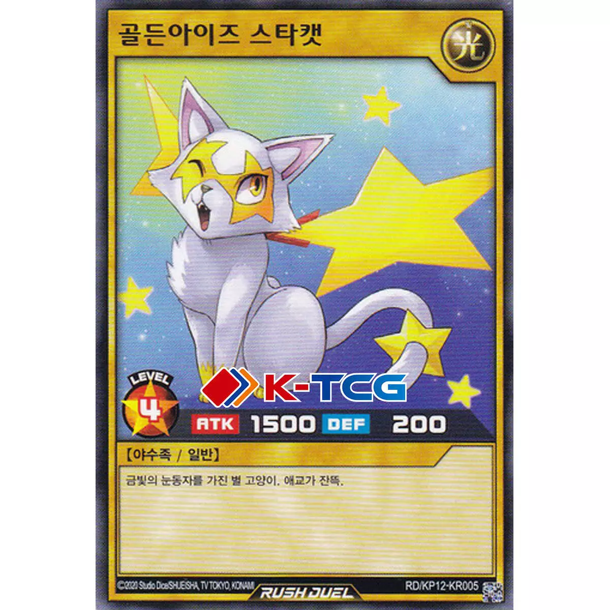 Yugioh Card "Golden-Eyes Star Cat" RD/KP12-KR005 Korean Ver Common13 março 2025
 -
 Ars no Kyojuu - Episódio 1 - Animes Online13 março 2025
 -
 Chainsaw Man Pack 213 março 2025
 -
 Manchester City ALL KITS, DREAM LEAGUE SOCCER, 2017, 2018, 2019, 2020, 2021, by TechiApkWorld13 março 2025
 -
 Anime Fairy Tail HD Wallpaper13 março 2025
 -
Comunidade Steam :: Vampire: The Masquerade - Bloodlines13 março 2025
-
 Shrek by futdiversoesrj on DeviantArt13 março 2025
 -
 Fallen vhs sans pixel art13 março 2025
 -
 Bom dia! – Liege Barbalho13 março 2025
